


Osteoarthritis of the hip (ATS) is a slowly destructive disease. Under the influence of a number of reasons, during the development of the disease, irreversible changes in the structure and properties of the hyaline cartilage occur, which lead to increased pressure on the joint surface and deformation or fusion. most of them. Given that mechanical overload is considered to be one of the main causes leading to the development of the disease, the hip joint is often affected by degenerative joint disease.
Anatomical features of the hip joint
The hip joint (TC) is the junction of the pelvis and femur. This articulation allows you to contract and stretch your lower limbs, raise your legs and pull them toward your body, and perform walking movements. From birth and throughout life, a person is subjected to a high load on the hip joint.
From the pelvic side, the "acetabulum" cavity joins the articulation, from the femur side, its bony end. Acetabulum has a collagen lip along the edges, which acts as a kind of cushion that holds the femoral head firmly in its recess. The cavity in the center of the acetabulum is covered with a collagenous membrane and is the site of attachment of the ligaments of the femur.
The composition of TS capsules includes ligaments:
- femur-pelvis - the strongest ligament that can withstand loads in excess of 200 kg and prevents excessive backward arching of the hips;
- thigh-pubic - responsible for the abduction and reduction of the thigh, thereby limiting its circular movement;
- femur - protects the car from shocks, reduces the load when riding and running;
- circle (ring) - prevents dislocation and holds the femoral head in the cavity of the pelvic cavity and is the basis of the joint pocket.
Numerous muscle groups and tendons allow the vehicle to move around three axes:
- Vertical (vertical).
- Horizontal (horizontal, front).
- Sagittal (front-back).
Osteoarthritis can occur in both healthy joints and become a continuation of existing diseases of the musculoskeletal system.
What is this disease?
The transparent cartilage performs the functions of shock absorption and protection from damage to the joint surface. ATS is a developmental disease in which the structure of collagen cartilage fibers changes, which then leads to their fragmentation and destruction. Cartilage fragments, if they enter the joint cavity, can cause inflammation. Bare surfaces undergo changes in bone tissue due to friction and increased pressure. The remaining cartilaginous tissue along the edges of the bony head grows to compensate with subsequent ossification, causing ankylosing spondylitis (immobilization of the bony junction). In the later stages, if not adequately treated, the patient completely loses mobility and becomes disabled. Destructive processes are stimulated for many reasons.
The following types of hip joints are available:
- Primary school. Its cause is not fully understood. Idiopathic (primary) osteoarthritis develops in a previously healthy joint. Usually, it develops in older people.
- Middle School. It is provoked by previous diseases of the articular apparatus, congenital abnormal development, changes in the work of vital organs and systems of human functioning.
The disease develops in one joint or affects both joints at the same time.
Cause of disease
Among the causes that contribute to the occurrence of the disease and its progression, the following are identified:
- Hereditary predisposition to the development of the disease.
- Musculoskeletal injuries (dislocations, fractures, sprains and dislocations).
- Irresistible systemic strength and physical activity.
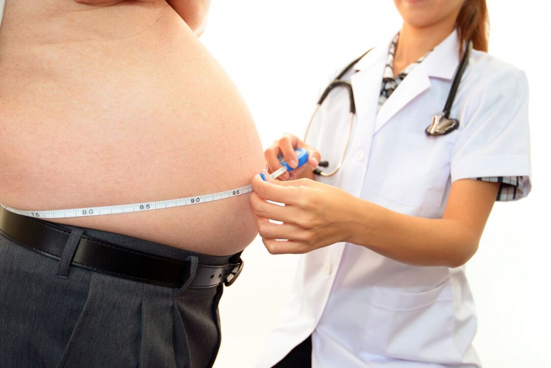
- Overweight.
- Dysfunction of the endocrine system (diabetes, psoriasis).
- Congenital pathologies of the structure and development of the musculoskeletal skeleton.
- Occupational characteristics of labor activities.
- Poor local traffic.
- Diseases previously caused by pathogenic flora.
- Legg-Calve-Perthes disease.
- Metabolic disorder (gout).
- No physical activity.
- immune disease.
These reasons may not always cause ATS. Most often, the activation of pathological processes can be triggered by:
- increased stress and physical activity;
- constant overwork;
- hypothermia of the vehicle or the whole body;
- sudden heavy lifting;
- hormonal imbalance;
- radiation exposure.
Symptoms of the disease
Symptomatic manifestations of ATS are similar to those of arthropathy in other joints.
The main characteristic symptoms of this disease are considered:
- Stiffness in the morning or after a long period of immobility.
- Reduced range of motion, altered gait.
- Pain is first caused by mechanical or physical stress, then continuous.
- Appears creaking, creaking, and clicking with sudden movements.
- Pronounced lameness on the affected limb.
- Presence of contracture (passive restriction of movement).
- Narrowing or closing of joint space (X-ray sign).
The severity of the signs of hip osteoarthritis depends on how advanced the disease is and how responsive the patient's body is.

Stages of coxarthrosis
Depending on the clinical presentation, four stages of hip osteoarthritis can be distinguished:
- Grade 1 hip osteoarthritis has no obvious pain and other manifestations. In the difficult-to-diagnose stage, the disease can be detected using biochemical studies of transparent cartilage tissue and determination of insufficient glycosaminoglycans. Patients experience joint pain and rarely pain with the onset of physical activity.
- Second degree hip osteoarthritis is characterized by changes in the density and elasticity of the cartilage fibers. Cracks and breaks appear. Depreciation function is reduced. The pain increases, radiates down to the groin area, and contracture movements of the affected limb are limited.
- At grade 3, the stratification of the cartilaginous fibers takes place with greater intensity. Joint surfaces are subjected to excessive pressure, ischemic foci develop. Cartilaginous tissue grows along the edge of the bone head. Pain at the damaged bone junction does not depend on the state of life and rest. With any movement, the joint has a creaking and creaking sound. Range of motion is reduced on all axes.
- The fourth degree is characterized by contact of the surfaces of the joint components with the formation of ulcers and indentations. The articular end of the femur is poorly fixed in the acetabulum, which leads to a violation of the comparison and separation of the articular surfaces. During this stage, the patient experiences severe pain due to narrowing, sometimes closing of the joint lumen and compression of nerve fiber bundles and blood vessels. Movement is limited, sometimes completely.
The classification of pathological changes caused by ATS is necessary to understand the mechanism and characteristics of the development of the disease. Determining the severity of the disease helps to determine the correct treatment and disability tactics (in the case of severe illness).
Possible consequences
The progression of ATS leads not only to deformation of the femoral head and pelvic cavity, but also to the development of pathological processes throughout the functioning of the articular apparatus.
Complications of hip osteoarthritis:
- synovitis (inflammation of the synovial membrane of a joint);
- aseptic necrosis of the femoral head;
- joint destruction (osteonecrosis);
- bursitis with a change in the amount of joint fluid;
- partial or total stiffness (immobilization of the joints);
- contractures (limited movement and inability to flex and extend the limb).
The development of ATS complications always leads to a worsening of the patient's general condition, quality of life, and loss of mobility without assistance.
diagnostic method
Diagnosis of hip osteoarthritis in the early stages is difficult. Symptomatic manifestations become noticeable only when bone and nerve fibers are involved in the pathological process.
During the medical examination in advanced stages, the following are noted:
- change the image in the matching border;
- painful palpation;
- sometimes flaccid tissues around joints;
- shorten the sick limb.
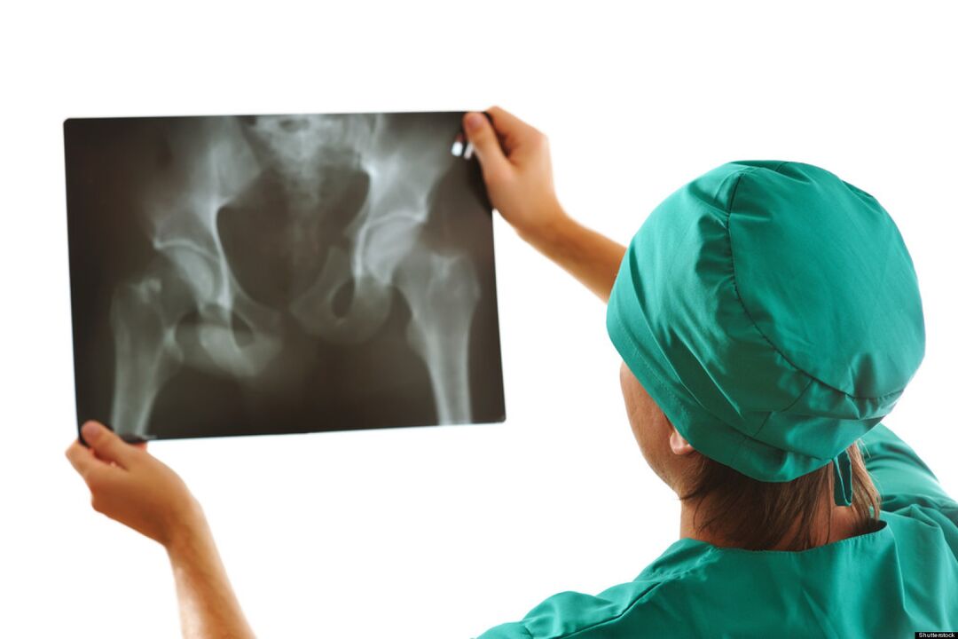
The main role in the diagnosis of ATS is assigned to the radiological examination. As an auxiliary diagnostic method used:
- Ultrasound, magnetic resonance imaging.
- CT scan.
- Perforation lubricates the joint (synovial fluid).
- Diagnosis by arthroscope (microprobe).
- Laboratory and biochemical tests of urine and blood.
Timely diagnosis improves the treatment prognosis and later life of the patient.
How to apply for disability?
It is not possible to completely cure this disease. To confirm your right to social assistance and to assign a disability group after passing the examination of narrow specialists, you must contact your doctor.

Indications for the determination of disability in case of hip joint are:
- oligoarthrosis (injury to no more than 2 joints) TS 2 degrees;
- combined knee osteoarthritis and grade 3 knee osteoarthritis;
- reduce the length of the affected limb by more than 6 cm;
- Phone exchange automatically flows response, is recorded.
In identifying disability groups will help:
- carefully collected anamnesis;
- conclusions of the medical advisory committee (MCC);
- diagnostic research results;
- through the committee of health and social experts (MSEC).
If the expert committee's decision is negative, it can be appealed to the higher authorities.
Prevent
Preventive measures are an easy way to avoid the development of this disease. Preventive measures include:
- Adhere to an active lifestyle.
- Control the indicators of body weight.
- Optimizing nutrition and mode of work and rest.
- Reduce mechanical and physical loads.
- Treatment of viral and infectious diseases.
- Prevention and prevention of injuries at home and at work.
- Regular preventive check-ups.
Inference
The answer to the frequently asked question: "Is there a cure for osteoarthritis of the hip? " Experts give a negative answer. The damaged cartilage tissue cannot be completely restored, nor can the deformation and destruction of the bones included in the joint be completely corrected. Do not ignore the minor manifestations of degenerative hip disease, which reduces the ability to prevent further development of the disease.